Erythrocyte Sedimentation Rate (ESR)
Erythrocyte sedimentation rate (ESR)
What is an Erythrocyte Sedimentation Rate (ESR)?
An erythrocyte sedimentation rate (ESR) is a blood test that that can show if you have inflammation in your body. Inflammation is your immune system's response to injury, infection, and many types of conditions, including immune system disorders, certain cancers, and blood disorders.
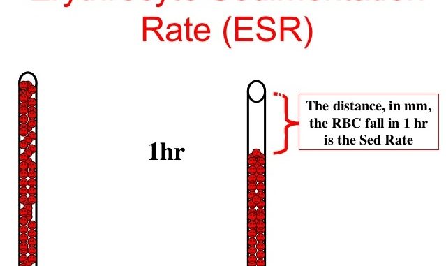
Erythrocytes are red blood cells. To do an ESR test, a sample of your blood is sent to a lab. A health care professional places the sample in a tall, thin test tube and measures how quickly the red blood cells settle or sink to the bottom of the tube. Normally, red blood cells sink slowly. But inflammation makes red blood cells stick together in clumps. These clumps of cells are heavier than single cells, so they sink faster.
If an ESR test shows that your red blood cells sink faster than normal, it may mean you have a medical condition causing inflammation. The speed of your test result is a sign of how much inflammation you have. Faster ESR rates mean higher levels of inflammation. But an ESR test alone cannot diagnose what condition is causing the inflammation.
Other names: ESR, SED rate sedimentation rate; Westergren sedimentation rate
What is it used for?
An ESR test can be used with other tests to help diagnose conditions that cause inflammation. It can also be used to help monitor these conditions. Many types of conditions cause inflammation, including arthritis, vasculitis, infection, and inflammatory bowel disease. An ESR may also be used to monitor an existing condition.
Principle
When an anticoagulant is added to the blood and this well mixed venous blood is placed in a vertical tube, erythrocytes tend to settle towards bottom leaving clear plasma on top. This rate of sedimentation of red blood cells in a given interval of time is called erythrocyte sedimentation rate (ESR).
As the erythrocytes sediments, in a period of one hours, 3 stages can be observed.
- Stage I: first 10 minutes
- It is initial period of aggregation during which rouleaux are formed and the sediment rate is low
- Stage II: next 4o minutes
- It is a period of fast setting. Sedimentation occurs at a constant rate during this period
- Stage III: next 10 minute or more
- The sedimentation again slows as it is the final period of packing of cells at the bottom of the tube
Sample
- Blood in EDTA or oxalate can be used.
Method for ESR estimation:
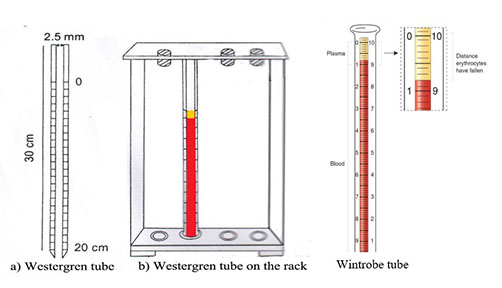
1. Westergren method
2. Wintrobe method
Materials required:
- Westergren tube or wintrobe tube
- Anticoagulant: 0.1 M sodium citrate
- ** in modified westergren method EDTA is used as anticoagulant
Procedure for ESR estimation:
- Withdraw 4 ml of venous blood
- Mix exact 10ml of sodium citrate with 4ml of venous blood in a tube
- Invert the tube 2-3 times to mix the blood thoroughly with anticoagulant
- Fill the westergren tube up to mark 0 and place in the rack at room temperature undisturbed and away from sunlight.
- Take the reading exactly after 1 hour. Record in millimeters from top surface of column to top of RBC sediments.
Result:
- Normal value of ESR
- Female:
- under 50 years- 20 mm/hr
- above 50 years- 30mm/hr
- Male:
- Under 50 years- 15mm/hr
- Above 50 years- 20 mm/hr
- Female:
Clinical application of ESR estimation:
- ESR test is non-specific test although it is used as indication of presence of disease
- ESR value increase during rheumatoid arthritis, chronic infection, carcinoma, tissue destruction and nephritis
- During pregnancy, ESR increase moderately from 10th or 12th weeks onwards and return to normal after delivery.
- ESR value decreases in sickle cell anemia and congestive heart failure (CHF).
Factors affecting ESR:
- There are several factors that affects sedimentation of erythrocytes.
Factors that increases ESR:
- Anemia:
- anemia increase ESR because the change in erythrocyte-plasma ratio favors rouleaux formation.
- Rouleaux is aggregation of RBCs together due to their discoid shape.
- Rouleaux have a decrease surface area and accelerate ESR
- Increase level of fibrinogen:
- it decreases the negative charge of erythrocyte, so RBC tend to remain apart and this promotes formation of rouleaux and increase ESR
- Immunoglobulin:
- increase antibody level in blood increase ESR
- Increase cholesterol level
- Rheumatoid arthritis
- Chronic infections
- Carcinoma
- Tissue destruction and other disease
- Anemia:
Factors that decrease ESR:
- Defibrinigenation:
- removal of fibrinogen decreases ESR
- Increase albumin and lecithin in blood
- Abnormal or sickle shape RBCs:
- abnormal or irregular shape of RBC lower ESR
- Congestive heart failure
- Defibrinigenation:
https://tmc.gov.in/tmh/PDF/Hemato%20Pathology%20Course/Rashida%20Ansari%20ESR.pdf
https://medicosage.com/erythrocyte-sedimentation-rate-esr-practical-method/
https://labpedia.net/erythrocyte-sedimentation-rate-esr-solution-and/
https://medlineplus.gov/lab-tests/erythrocyte-sedimentation-rate-esr/
https://www.medicine.mcgill.ca/physio/vlab/bloodlab/ESR.htm
.
.
.
.
.
Comments
Post a Comment